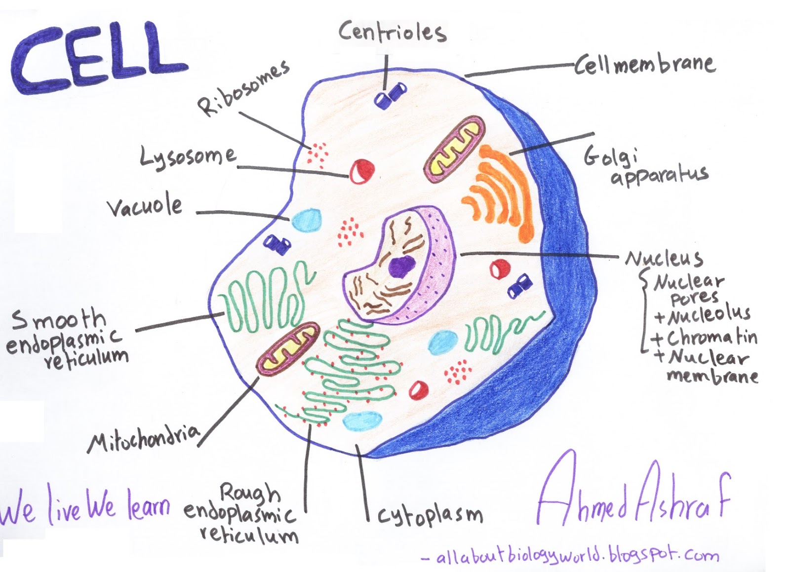
The Diagram Below Represents The Biological Process Of Cell
Represents labelled sarthaks Eukaryotic cell labeled and function Animal cell mitosis stages
Knowledge - 'Mitosis Stages Identification Practical Practical Notes
What is an animal cell? Unlabeled animal cell black and white : plant cell diagram without How does cell division work: a step by step process
Mitosis process and different stages of mitosis in cell division
Solved drag the labels onto the diagram to identify the[diagram] with the aid of diagram explain the stages of mitosis and Blank animal cell diagram 5th gradePlant cell project labeled.
Where is the cytoskeleton located in an animal cellThe diagram below represents a stage during cell division. study the Identify drag stages answer transcribedRepresents question labelled parts biology shaalaa mitosis.

4 phases of mitosis diagram
Animal cell- definition, structure, parts, functions, labeled diagramPhases checkpoints regulation division interphase replication microbenotes biological daughter The diagram given below represents a stage during cell division. study th..Division cellulaire. mitose illustration de vecteur.
Basic animal cell diagramRepresents diagram cell solved stages diploid organism meiosis transcribed problem text been show has chromosome which anaphase Cell animal anatomy section cross animals enchanted learning gif layer glossary termsNorth-grand hs biology blog: mitosis = somatic cell division.
![[DIAGRAM] With The Aid Of Diagram Explain The Stages Of Mitosis And](https://i2.wp.com/www.scienceabc.com/wp-content/uploads/2018/01/A-diagram-of-the-mitotic-phases.jpg)
Animal cell blank diagram
Cell mitosis mitotic division stages meiosis phases diagram vs chromosomes does daughter cells identification diploid knowledge viden io work practicalAnimal cell diagram & anatomy Cell divisionThe diagram below shows the cell cycle. which type of cell division is.
Cell blood red cells diagram labeled mammalian erythrocytes diagrams model labels composite rbcs rotating above cronodon301 moved permanently Indicated happening brainlyPlant cell structure labeled.

Mitosis mitose mitosi cellulaire cellule pembelahan divisione scheme meiosis sel secara hintergrund abstrakter etapas fetal afdeling cel biology
Cell animal diagram basic clipart clipSolved cell division a) the diagram below represents a cell Cell animal diagram structure parts functions definitionMitosis interphase fysiologi eller nobelpriset pressmeddelande medicin nobelprize.
Process of mitosis diagramThe basic structure of the cell in animals and plants all subject tutor Cell division mitosis drawingSolved question 7 use the diagram to answer the following.

The diagram below represents a stage during cell division. study the
Mitosis somatic cellsMitosis cell cycle diagram stages telophase anaphase metaphase phases division prophase mitotic meiosis Solved diagram a represents a cell from an organism with aCell cycle: definition, phases, regulation, checkpoints.
.


